


A connected recorder can still produce an unusable trace. Assess wireless ECG signal quality by checking electrode contact, waveform fidelity, filtering, and data continuity against the intended examination.
A Bluetooth connection icon tells you about the link. It doesn’t establish whether the electrocardiogram (ECG) preserves the information your clinical team needs. Nor does a smooth line prove that every recorded sample reached the saved file. Those questions need separate evidence.
If you’re choosing equipment for a clinic, lab, or distributor, a polished demo is only a starting point. You need to know which device, software, cables, and settings produced the result. Otherwise, two suppliers can appear to demonstrate the same capability while testing different conditions.
This guide helps you separate noise at capture from data-transfer problems, understand quality scores, and plan an equipment review. It draws on professional ECG guidance, an established noise-test database, and FDA information about wireless medical devices. The focus is equipment review for professional use. Qualified staff make clinical judgments.
Always follow the selected system’s current instructions for use and your organization’s approved procedures.
Key takeaways
- Check three stages separately: skin to recorder, recorder to application, and application to saved record.
- Electrode motion, muscle activity, and baseline drift can affect a trace even when Bluetooth remains connected.
- A signal quality index needs a defined task, method, and denominator; it isn’t a diagnosis.
- Compare filtering, missing-data handling, and exported records on the exact proposed hardware and software configuration.
What determines wireless ECG signal quality?
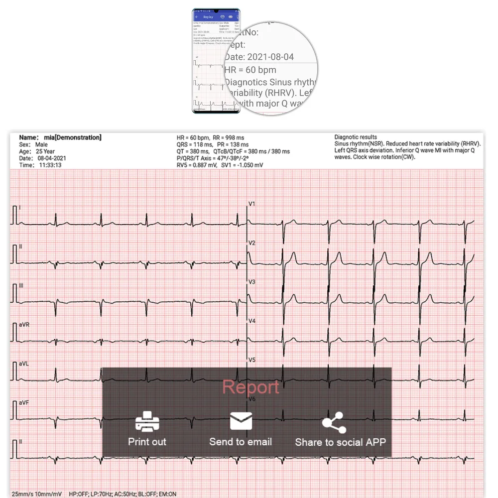
Useful quality means the recording supports its intended task. Counting beats, reviewing rhythm, and assessing detailed waveform shape place different demands on the data. Start with the examination your service provides, then ask which evidence supports that use.
Think of the recording as three stages. First, electrodes and the acquisition hardware capture a small electrical signal. Next, the wireless link transfers data to an application. Finally, software displays, processes, stores, and exports the recording.
A fault can arise at any stage. Poor skin contact can disturb the captured signal. Link problems can interrupt data transfer. Display or export settings can change what a reviewer sees.
Calling every problem “Bluetooth noise” makes it harder to identify the cause.
For a model-specific starting point, review Vales and Hills Biomedical Tech. Ltd. (V&H)’s Android iCV200 product information. The listing describes Bluetooth acquisition with the aECG application; confirm the current configuration before planning a demonstration.
Hypothetical example: Alex, a biomedical engineer, reviews two recordings during an equipment demonstration. Both show an active connection, but one has a drifting baseline while the other contains a marked missing interval. Alex records these as separate issues and asks the supplier to investigate each under an approved test plan. The outcome is a clearer fault report, rather than a single complaint that the wireless system is unreliable.
This example describes a review method, not a V&H test result.
Trace the source of noise before changing settings
Begin with contact, movement, and lead configuration
ECG motion artifact can begin where an electrode meets the skin. Movement changes contact conditions, and pulling on a patient cable can disturb that interface. Muscle activity can also add electrical activity that overlaps the ECG. A wireless recorder still needs its documented patient connection; wireless data transfer doesn’t automatically remove electrode leads.
ECG baseline wander describes a slow shift in the trace’s baseline. Breathing, movement, and unstable contact are possible contributors. A trained operator should review preparation, electrode condition, placement, and cable support using the current instructions. Avoid adopting an unverified preparation method simply because it improved one demonstration.
Placement matters as well as contact. A conventional 12-lead ECG uses 10 electrodes to obtain different electrical views of the heart. Moving an electrode changes the view being recorded. It isn’t a general-purpose correction for a noisy trace, and changes can affect comparisons with earlier recordings.
The 2017 ANMCO/AIIC/SIT consensus document on ECG signal quality discusses contact, placement, acquisition, processing, and display as parts of the same quality problem. Its recommendations concern defined applications. They don’t establish one preparation method or filter setting for every device.
Treat appearance as a clue, not proof of the cause
A waveform’s shape can guide a fault check, but it cannot prove the cause. Similar-looking problems can have different causes. Keep the original record and settings before changing the setup.
| Observation | Possible area to investigate | What to record |
|---|---|---|
| Slow baseline drift | Contact, movement, breathing, or processing | Lead affected, activity, electrode condition, filter settings |
| Dense fine interference | Muscle activity or electrical interference | Test environment, activity, affected leads, settings |
| Flat-topped or truncated signal | Input overload, display scale, or processing | Original data if available, gain, input conditions |
| Gap, frozen display, or discontinuous time | Acquisition, transfer, application, or storage | Timestamp, connection messages, logs, saved record |
This table supports technical investigation, not diagnosis. A flat segment, for example, must not be assumed to represent either a patient event or a wireless failure without assessment. Follow clinical escalation procedures when a patient recording is uncertain.
Measure wireless ECG signal quality for the intended task
Review waveform fidelity alongside visual clarity
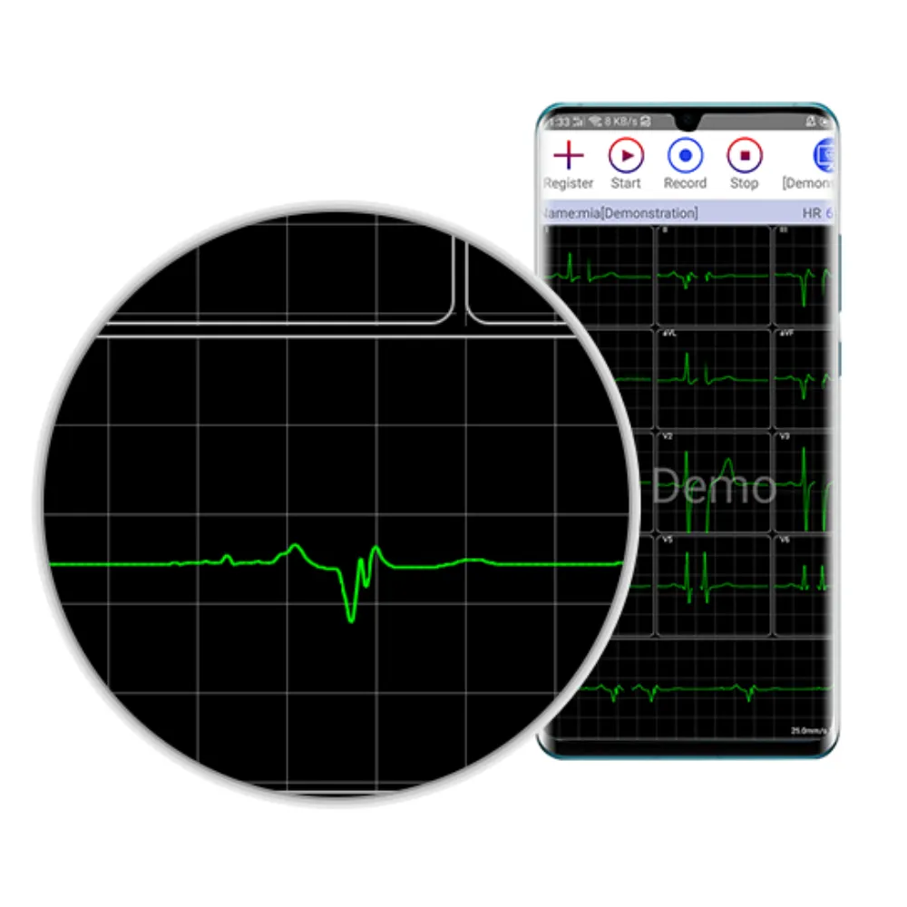
Waveform fidelity means preserving the signal features needed for the examination. The relevant question isn’t simply whether the line looks clean. Ask whether processing changes the shape, timing, or amplitude that qualified reviewers need to assess.
Filters can reduce unwanted components, but their effect depends on their design and settings. A setting that suppresses baseline movement can also affect low-frequency waveform features. Removing high-frequency content can alter sharp deflections. The consensus document explains why a visually stable trace can still lose useful information.
Request the documented sampling rate, frequency response, filter options, and test conditions for the selected configuration. Sampling rate describes how frequently the signal is measured; bandwidth describes the frequencies a system preserves. Neither figure alone proves diagnostic performance. Ask which settings affect the displayed trace, stored samples, measurements, and printed report.
Where the system supports access to less-processed data, ask qualified staff to compare that view with the processed output. If it doesn’t provide that access, request an explanation of the processing and supporting verification. Don’t assume a PDF is a copy of every acquired sample.
Understand quality indexes and usable coverage
An ECG signal quality index uses a defined method to score selected traits of a recording. One score might show how closely two beat detectors agree. Another might rate noise, shape, or missing samples.
A high score answers the question that method was built to ask. It doesn’t prove that every waveform feature is preserved.
Ask what the score means, how much data each window covers, and where the pass point came from. Ask which groups it was tested on. Find out whether rejected segments remain visible to reviewers.
A report based only on clean sections can hide how much data was left out. Ask for the total time used in the calculation.
For continuous recordings, a useful review can separate three durations: the time recording was intended, the time data was available, and the time judged usable for the stated analysis. Define how overlapping windows, disconnected periods, and partially acceptable leads are counted. There is no universal acceptance percentage for all ECG applications.
For a brief resting examination, the relevant question may instead be whether the required interval contains acceptable data across the required leads. A long-duration coverage percentage can hide a short but important interruption. Match the measure to the examination rather than borrowing the easiest metric from a brochure.
Keep test-dataset numbers in context
The PhysioNet MIT-BIH Noise Stress Test Database separates baseline wander, muscle artifact, and electrode-motion noise. It contains 12 half-hour ECG records and three half-hour noise records. Its controlled noisy ECGs use six signal-to-noise ratio levels, from 24 to -6 decibels.
Signal-to-noise ratio compares signal and noise under a defined calculation. In controlled test data, the added noise is known. In a patient recording, separating true cardiac content from artifact is harder. A quoted SNR needs its calculation, signal source, bandwidth, and test conditions before it becomes a meaningful comparison.
PhysioNet also notes that electrode-motion artifact can resemble ectopic beats and resist simple filtering. That is a reason to review the source signal and intended analysis carefully. The database’s noise levels are algorithm-test conditions, not clinical pass thresholds or evidence about a V&H product.
Test transmission and the saved record separately
Use a documented, approved evaluation plan
A wireless ECG testing plan should list the device, app version, phone or tablet, operating system, patient cable, and recording mode. Keep that setup fixed and change one factor at a time. A change in software or cables can make an earlier test hard to repeat.
For engineering checks, use a compatible ECG simulator where the manufacturer and your technical team permit it. A known input helps you check the device without changes in a patient signal. A bench test does not prove clinical validity. It can help you check timing, channels, reports, and what happens when a link breaks.
Test within permitted operating conditions and the facility’s approved process. Don’t introduce radio interference or alter clinical equipment to create a fault. If a controlled interruption test is needed, agree on a safe bench method with the supplier and responsible technical staff.
The FDA’s wireless medical device information identifies coexistence, quality of service, security, and electromagnetic compatibility as areas to consider. Nearby wireless activity can disrupt communication. This supports evaluating the intended environment; it doesn’t establish a guaranteed range or a universal Bluetooth ECG data loss rate.
Compare acquisition, live display, and export
Ask what happens when the link is interrupted:
- Does acquisition stop?
- Is data stored locally?
- Can anything be recovered later?
- How is the missing interval identified?
These are model-specific questions. Don’t assume a recorder has buffering or recovery because a different product does.
Record elapsed time, available timestamps, any warning messages, and the interval affected. Then inspect the stored recording and required export with qualified staff. A display freeze doesn’t by itself prove that acquisition stopped, while a resumed display doesn’t prove that the missing interval was recovered.
Hypothetical example: Morgan, a technical evaluator, runs a supplier-approved bench test using a simulator and a supported tablet. They note a display pause at an elapsed time of 40 seconds, then compare the application record with its exported report. The team cannot resolve the difference from the report alone, so Morgan saves the test settings and asks for the documented storage behavior. The outcome is a reproducible support request, not an assumed diagnosis of packet loss or a claimed product defect.
Match the review to the platform: Compare V&H’s Android wireless ECG options and iOS wireless ECG options, then confirm the exact recorder, application, and supported versions for the proposal. Support for a platform alone doesn’t prove equal quality or the same functions.
Ask for evidence that matches the proposed configuration
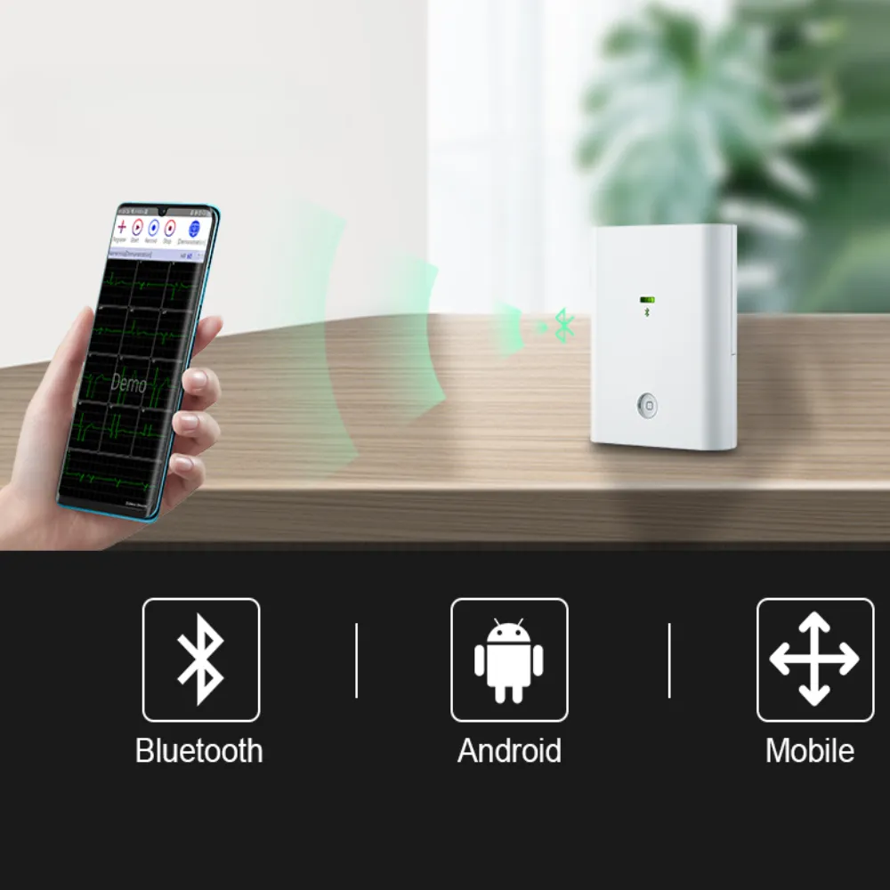
A useful supplier response ties each claim to its test conditions. “High-quality signal” is incomplete without the model, accessories, application, recording mode, and evaluation method. Ask for documents that your clinical and technical teams can assess together.
Start with the current instructions for use and the supported hardware/software list. Then request the documented acquisition specifications, filter behavior, and any relevant verification reports. If a report covers a different device version or app, ask whether it applies to the proposed setup.
For V&H, the Android iCV200 listing identifies Bluetooth, the aECG application, and 12-lead acquisition. Those facts help define an inquiry. They don’t establish a measured noise level, uninterrupted transmission, or a particular diagnostic accuracy. Obtain current model documentation for those questions.
Use a requirements table to keep the evidence review concrete:
| Requirement | Evidence to request | Decision owner |
|---|---|---|
| Required examination and lead configuration | Intended-use documentation and model specification | Clinical lead |
| Waveform processing and fidelity | Filter descriptions and applicable verification | Clinical and biomedical teams |
| Continuity and interruption behavior | Documented behavior and approved bench demonstration | Biomedical and IT teams |
| Saved record and reporting | Representative nonpatient record, format, and settings | Clinical and records teams |
| Configuration and destination market | Exact bill of materials and applicable documents | Procurement and regulatory staff |
This is a planning template, not a set of universal pass criteria. Define acceptance conditions before the demonstration, including who can approve an exception. Keep unresolved questions visible instead of converting a supplier’s explanation into an unsupported guarantee.
Finally, agree on a practical escalation route for poor recordings. Ask what nonpatient test files, settings, and logs support requires. Keep identifiable patient data out of ordinary inquiry messages and use your organization’s approved handling process when clinical data is needed.
Questions about wireless ECG signal quality
Does Bluetooth make an ECG noisier?
Bluetooth describes data communication, not the quality of skin contact or the acquisition hardware. Wireless interference can disrupt transfer, while contact and muscle artifact can occur with a stable link. Evaluate both pathways rather than attributing every noisy trace to Bluetooth.
Can filters fix every poor trace?
No. Filters have limits and can change useful waveform content. Electrode-motion artifact is particularly difficult because it can resemble cardiac activity. Correct documented setup issues first, and have qualified staff judge whether a recording is suitable for its intended use.
Is a signal quality index a diagnosis?
No. A quality index evaluates selected signal properties using a defined method. It does not replace clinical interpretation. Request the method, threshold, and validation scope before using an index in an equipment comparison.
What if the live display and export differ?
Preserve the original recording and configuration details. Ask how the software processes, stores, and renders each output, including scale, filtering, timing, and missing intervals. Don’t treat either view as definitive until the difference is explained through the documented workflow.
Turn the quality review into a purchasing decision
Assess wireless ECG signal quality through a chain of evidence: correct acquisition, preserved waveform features, reliable transfer for the intended use, and a saved record that reviewers can interpret. A connection icon or an attractive demonstration covers only part of that chain. Use each observation to ask a specific question.
Bring the clinical, biomedical, IT, and procurement teams into the same review. Agree on the examination, the proposed configuration, the acceptance method, and the person responsible for each decision. Note any missing evidence beside the relevant requirement. That makes the result easier to explain when the purchase is reviewed later.
Request V&H model-specific ECG documentation with the recorder model, intended examination, preferred platform, and destination market. Include your questions about filtering, data continuity, and report handling, together with the documents your team needs to assess them. A clear request helps the supplier respond to the right questions. It also helps your team compare proposals on the same terms.
Post time: November 20, 2026


